Skip to main content
Search
Personal space
Space reserved for the UCLouvain community
E-mail or username
*
Password
*
Create new account
Request new password
Math question
*
2 + 1 =
Log in
FR
EN
Brain-inter-atlas
interactive atlas of neuroanatomy
Toggle navigation
Telencephalon
Internal configuration – white matter
Internal configuration – white matter
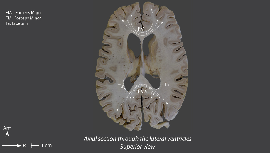
White matter
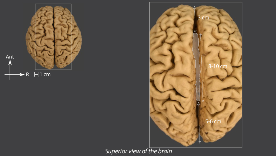
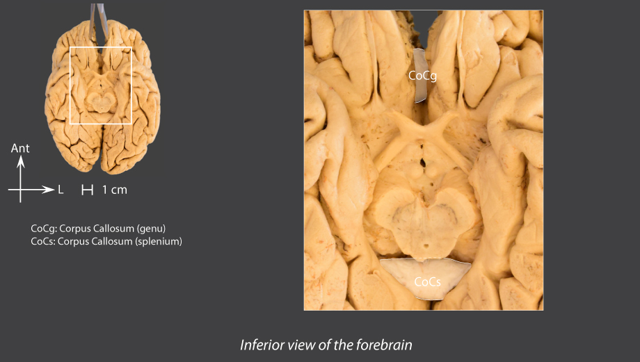
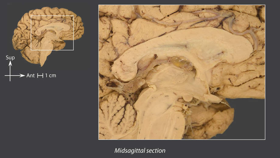
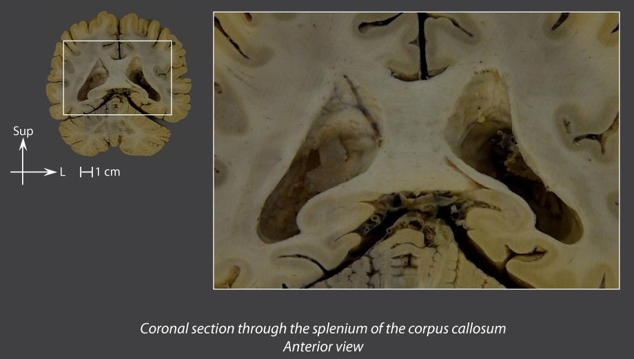
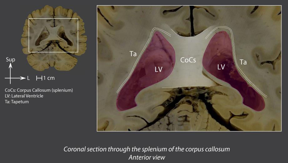
Corpus callosum
Arch-shaped bridge of white matter between the two cerebral hemispheres
Front end: rostrum, behind and underneath the genu of the corpus callosum
Back end: splenium
Anterior angles:
forceps minor
Posterior angles: bundle of fibres, divided into two parts:
Lateral part: tapetum, contributes to form the superior and lateral walls of the inferior and posterior horns of the lateral ventricle
Medial part:
forceps major
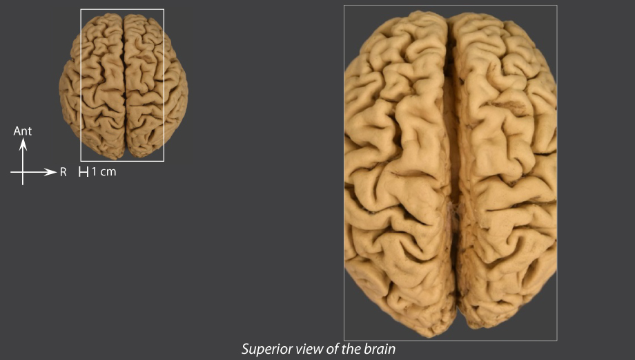
Superior view of the brain
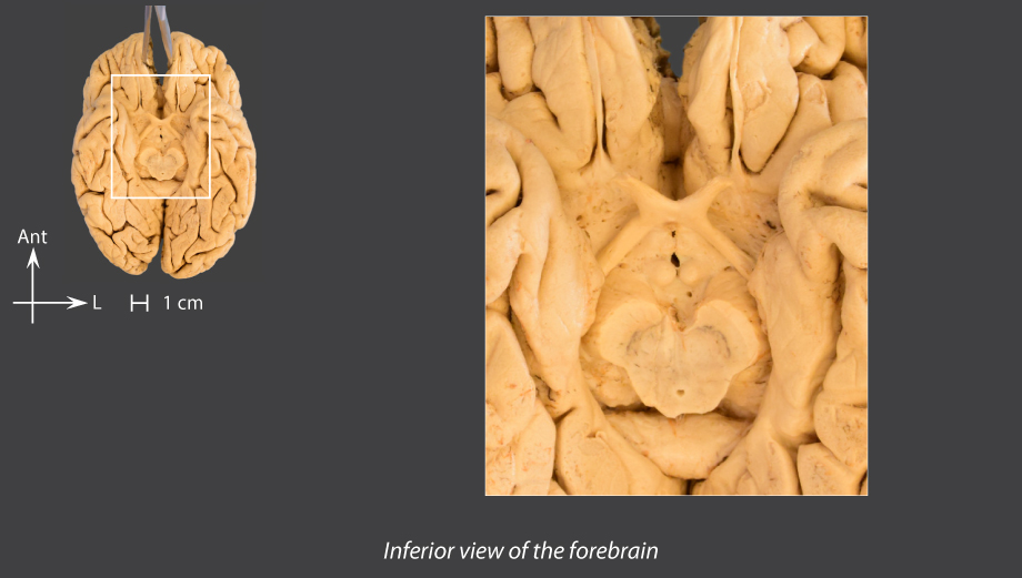
Inferior view of the forebrain
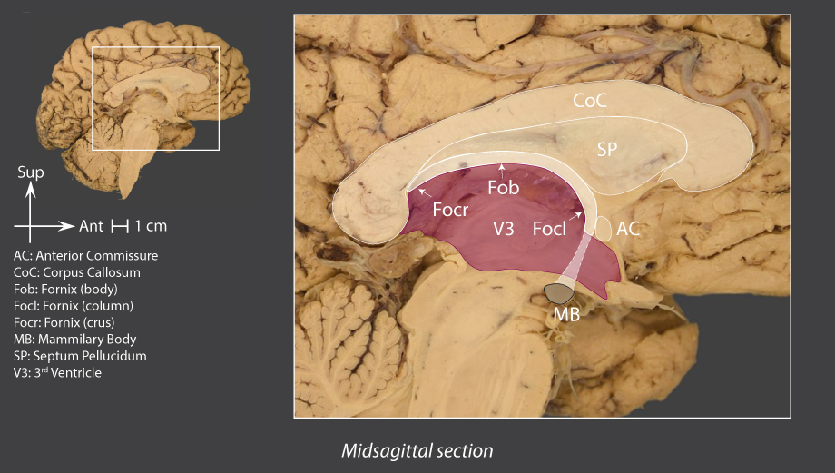
Midsagittal section
Coronal section through the splenium of the corpus callosum
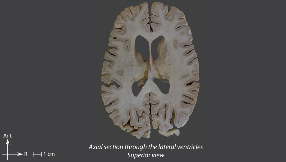
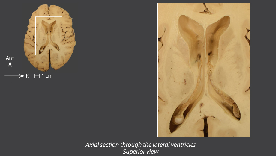
Axial section through the lateral ventricles
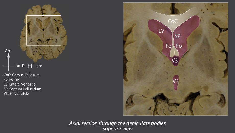
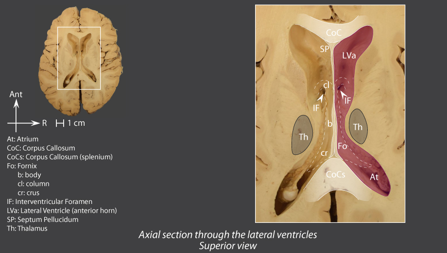
Septum pellucidum
Thin median and sagittal membrane
Separates the anterior horns of the lateral ventricles
Underneath the corpus callosum and above the
fornix
Midsagittal section
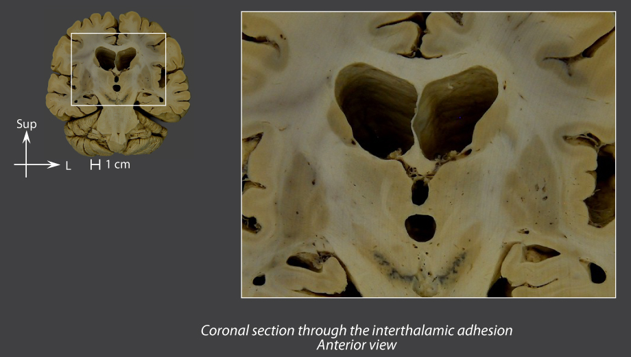
Coronal section through the interthalamic adhesion
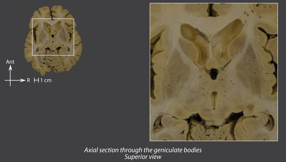
Axial section through the geniculate bodies
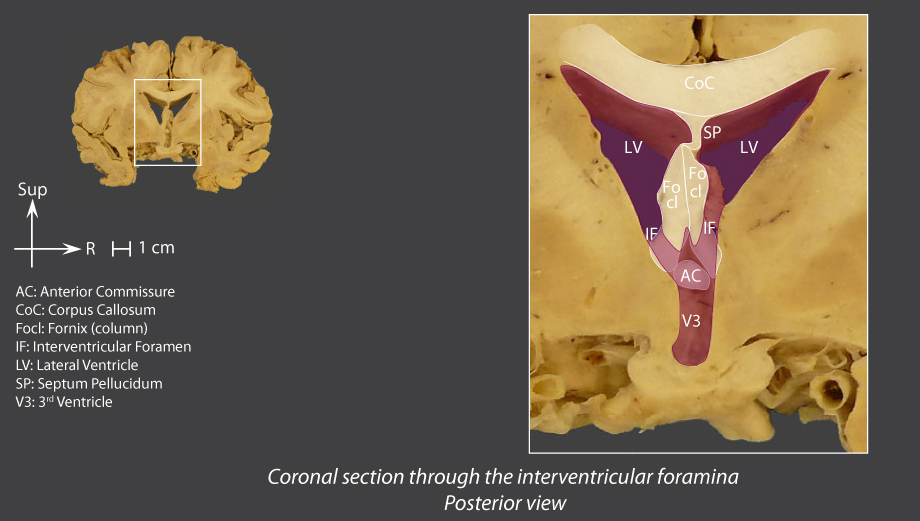
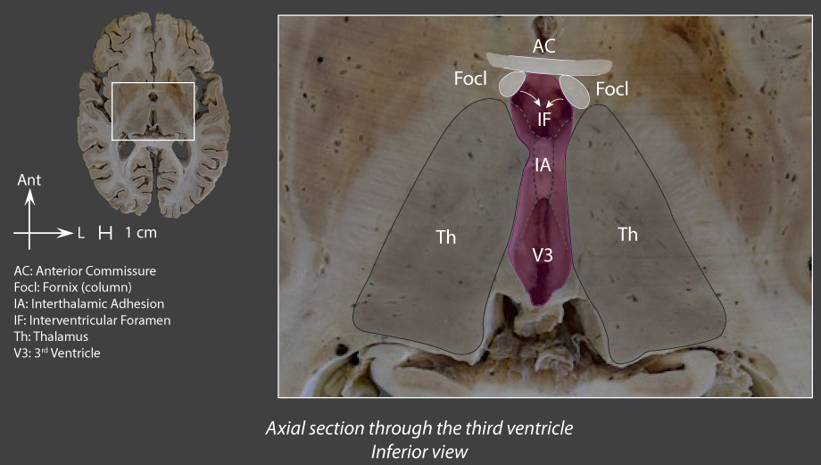
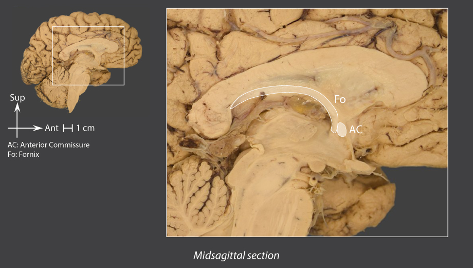
Fornix
Double cord of fibres above the thalami, underneath the corpus callosum
Forms part of the roof of the third ventricle
Three parts:
Crus of fornix
- Fibres from the alveus form the fimbria, then the crus of
fornix
- It converges medially and unites with the contralateral crus of
fornix
while adhering to the inferior surface or the corpus callosum
Body of fornix
- Linked to the contralateral body of
fornix
by the commissure of
fornix
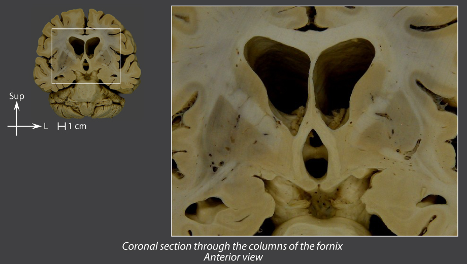
Column
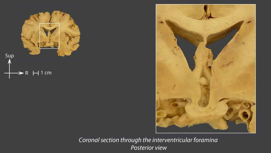
- Splits off from the contralateral column and contributes to form the interventricular foramen
- Extends into the mamillary body
Midsagittal section
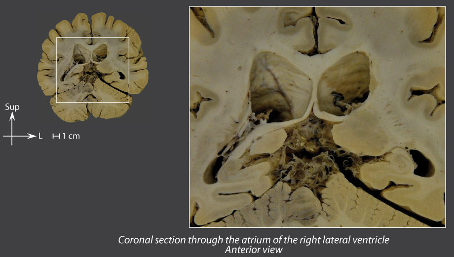
Coronal section through the atrium
Coronal section through the interthalamic adhesion
Coronal section through the columns of the fornix
Coronal section through the interventricular foramina
Axial section through the lateral ventricles
Axial section through the third ventricle
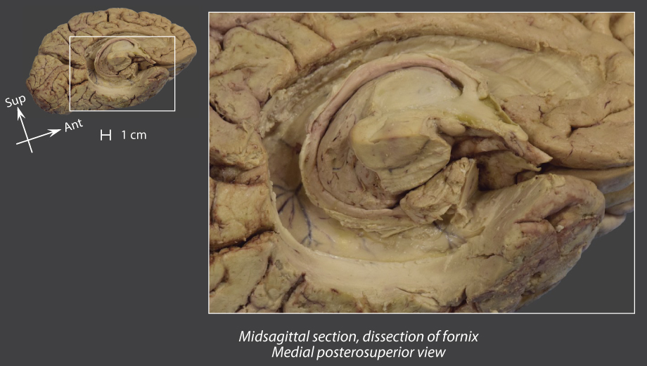
Midsagittal section, dissection of fornix
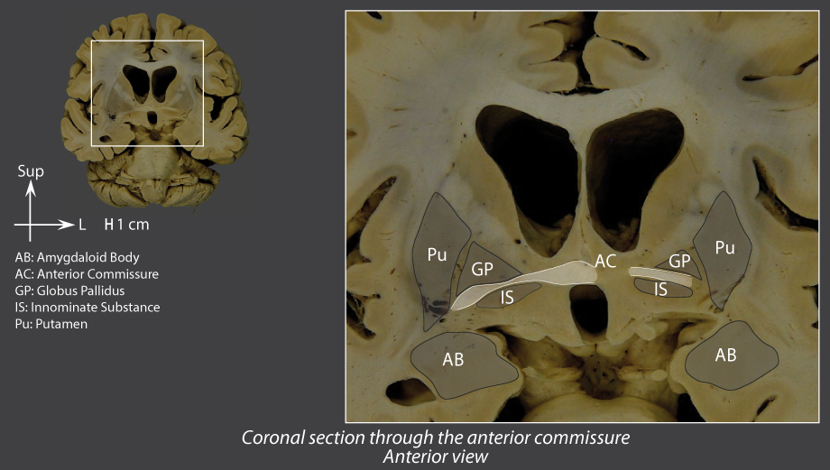
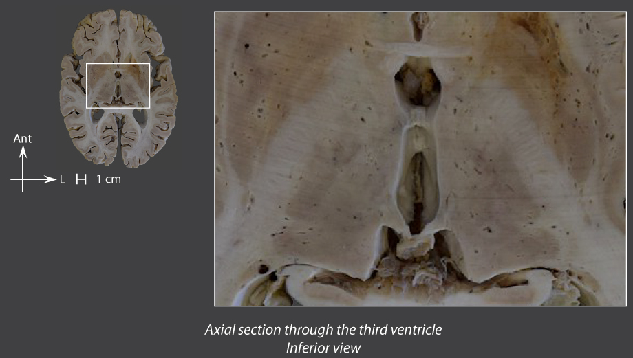
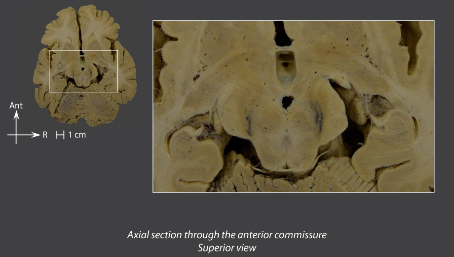
Anterior commissure
Thin cord that connects together the ventral parts of the cerebral hemispheres
Enables interconnections between the temporal lobes (cortex and amygdaloid bodies)
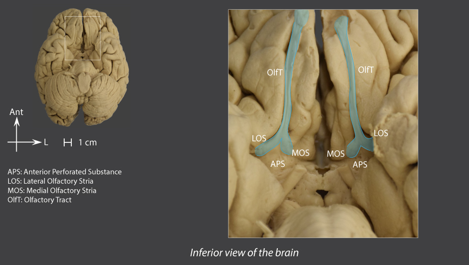
Contains fibres of the medial olfactory stria stemming from the olfactory tracts
Midsagittal section
Coronal section through the interventricular foramina
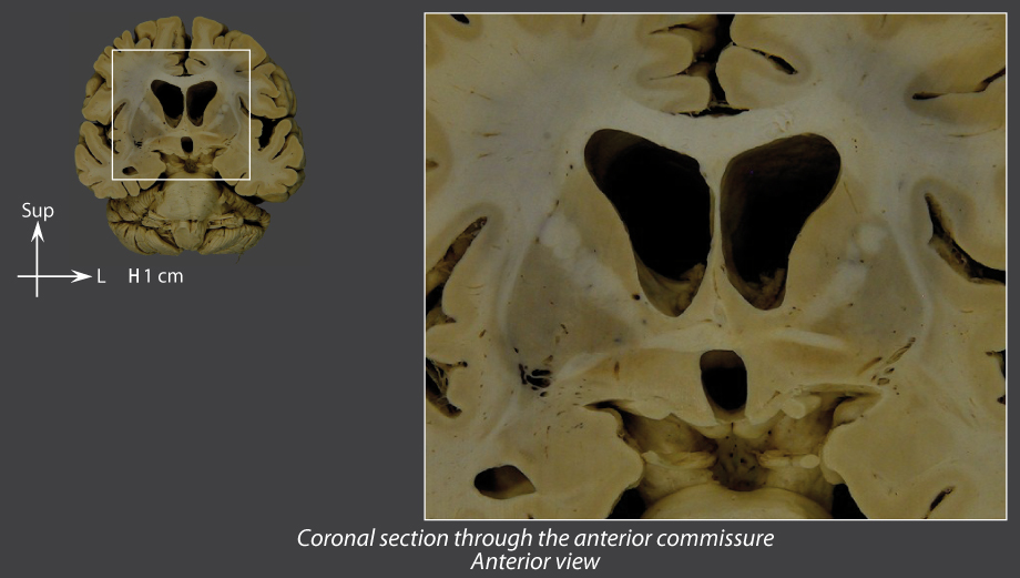
Coronal section through the anterior commissure
Axial section through the third ventricle
Axial section through the anterior commissure
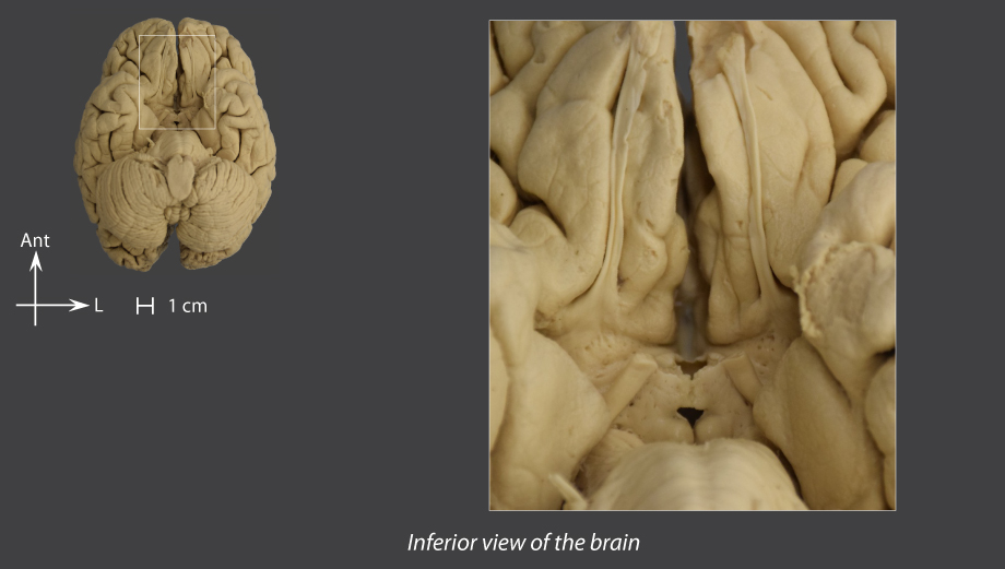
Inferior view of the brain
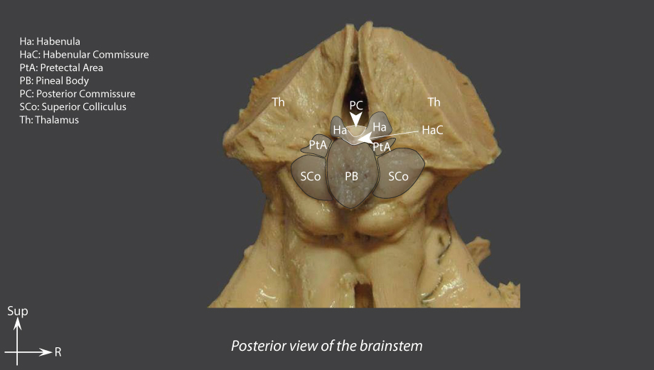
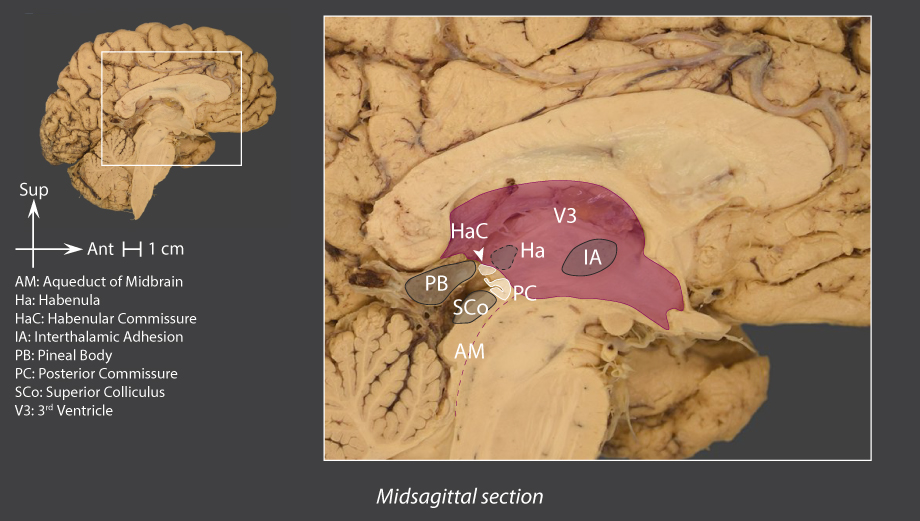
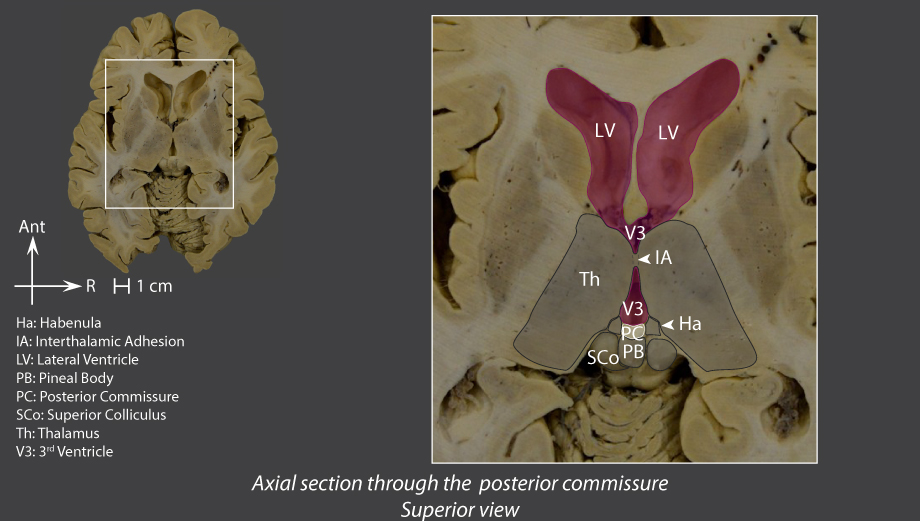
Posterior commissure
Thin bundle of fibres
Allows pretectal interconnections
Contains habenulotectal fibres
Posterior view of the brainstem
Midsagittal section
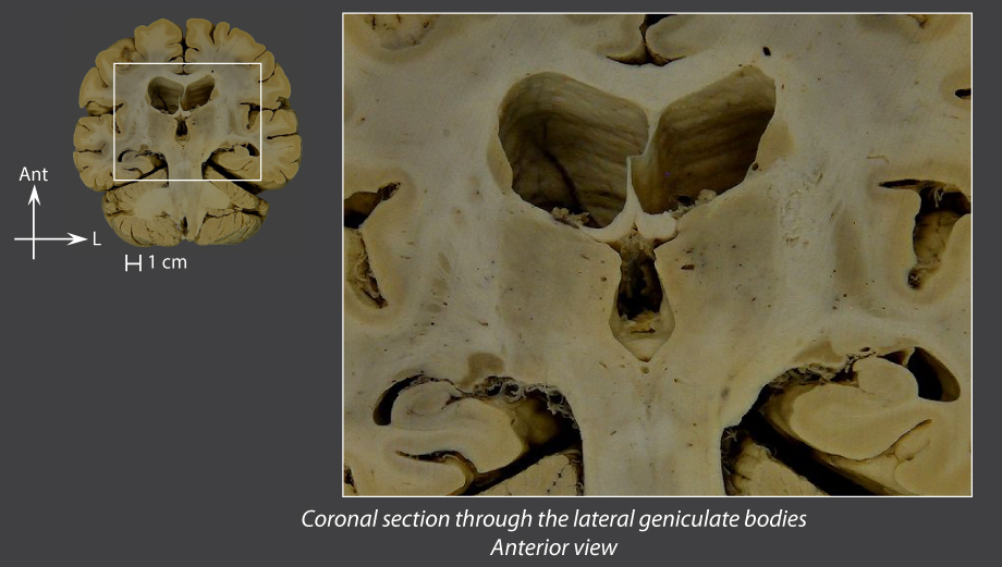
Coronal section through the lateral geniculate bodies
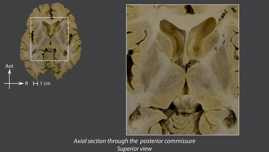
Axial section through the posterior commissure
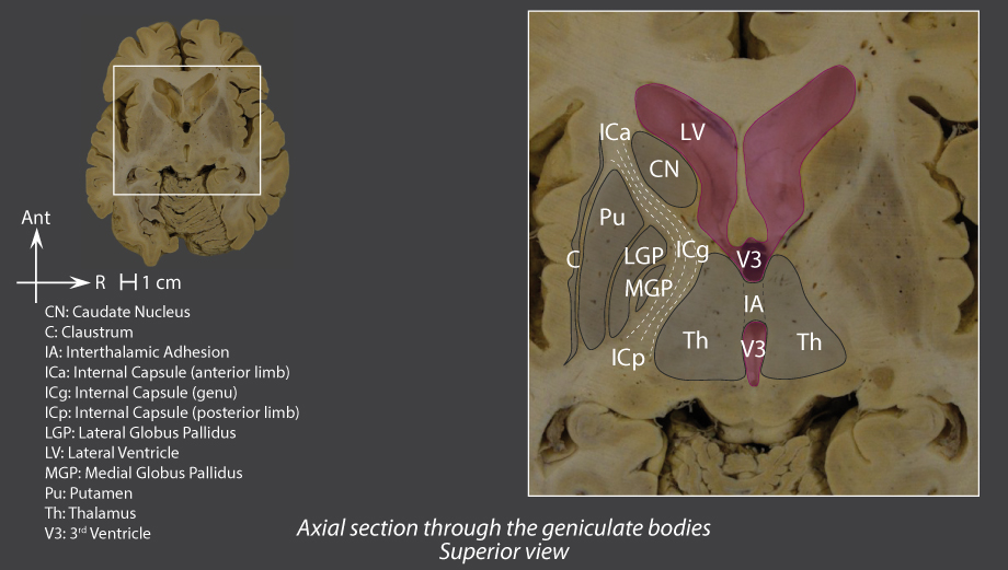
Internal capsule
Large conduction band that wanders along between the thalamus and the lenticular nucleus
Three sub-sections:
Anterior limb
Genu
Posterior limb
Downward and laterally-oriented
Extends into the cerebral peduncles
Axial section through the geniculate bodies
Coronal section through the interthalamic adhesion
Outline
Telencephalon
External configuration
Internal configuration – cerebral cortex
Internal configuration – hippocampus
Internal configuration – nuclei
Internal configuration – white matter
Schematic internal structure
Internal configuration – nuclei
Schematic internal structure
Introduction
Telencephalon
External configuration
Internal configuration – cerebral cortex
Internal configuration – hippocampus
Internal configuration – nuclei
Internal configuration – white matter
Schematic internal structure
Diencephalon
Midbrain
Cerebellum
Pons
Medulla oblongata
Spinal cord
Meninges
Vascularization
Ventricles of the brain
Sectional anatomy
Contributors
References
Menu