Skip to main content
Search
Personal space
Space reserved for the UCLouvain community
E-mail or username
*
Password
*
Create new account
Request new password
Math question
*
9 + 0 =
Log in
FR
EN
Brain-inter-atlas
interactive atlas of neuroanatomy
Toggle navigation
Telencephalon
Internal configuration – nuclei
Internal configuration – nuclei
Embryologic origin:
ventral pallium and subpallium
Classification of the nuclei of the telencephalon according to their embryologic origin
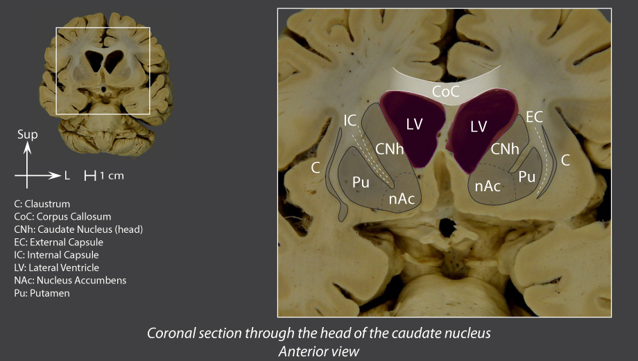
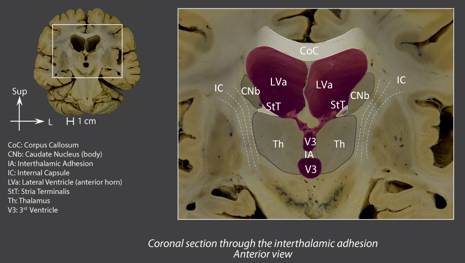
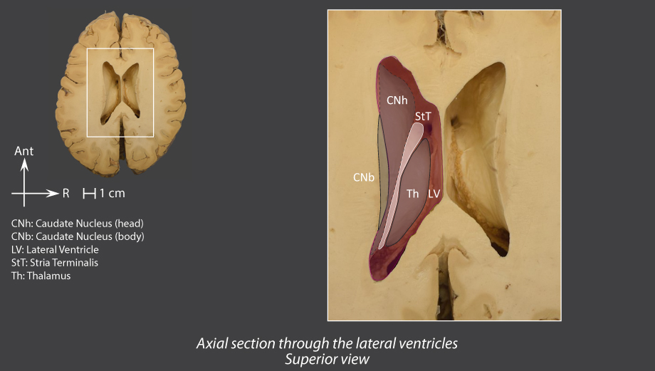
Caudate nucleus
Horseshoe-shaped, around the lateral ventricle and the thalamus
Three parts:
Head
Front: forms part of the floor of the
anterior horn of the lateral ventricle
Laterally: in continuity with the front end of the putamen, via the nucleus accumben
Above the anterior perforated substance
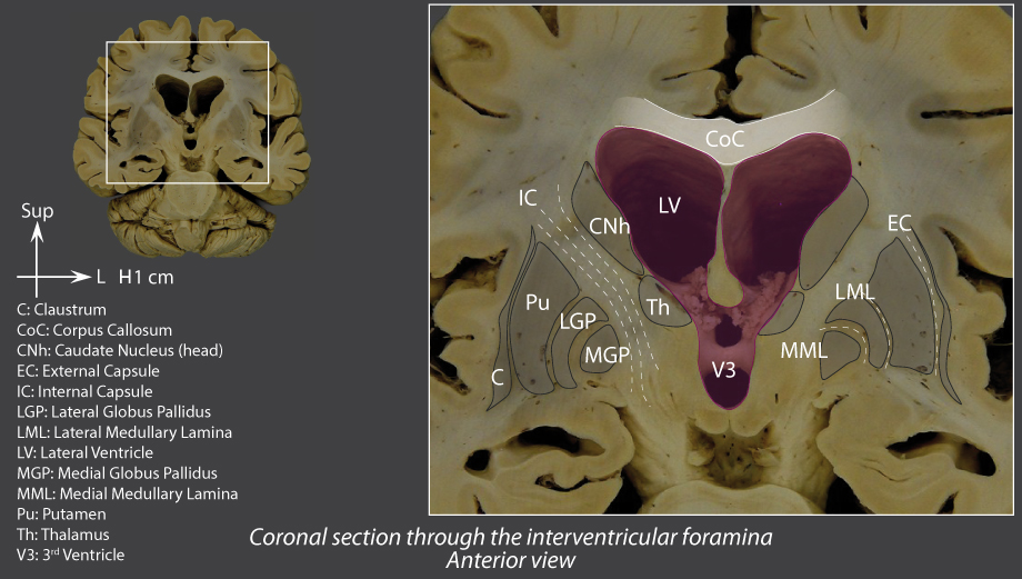
Body
Medially: forms part of the superolateral wall of the lateral ventricle
Laterally,from top to bottom: corpus callosum then internal capsule
Tail
Curves around the pulvinar before diving forward, along the roof of the
inferior horn of the lateral ventricle
Front: near the
amygdaloid body
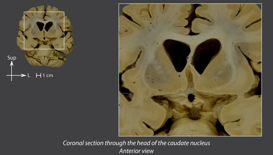
Coronal section through the head of the caudate nucleus
Coronal section through the interthalamic adhesion
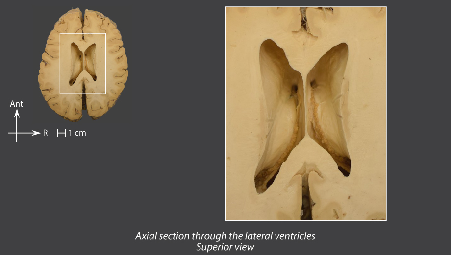
Axial section through the lateral ventricles
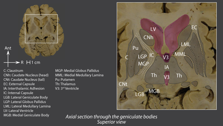
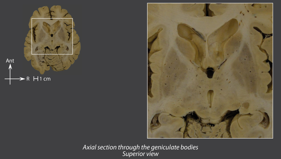
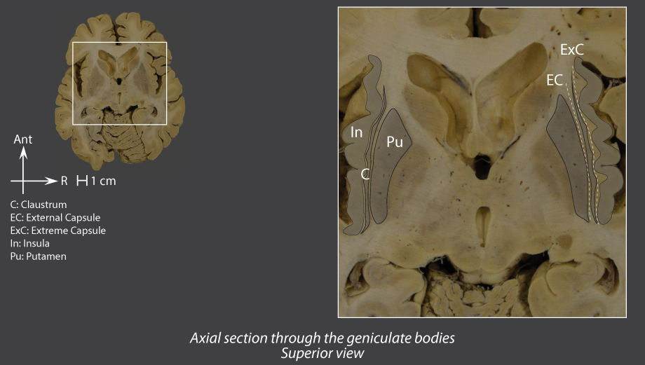
Axial section through the geniculate bodies
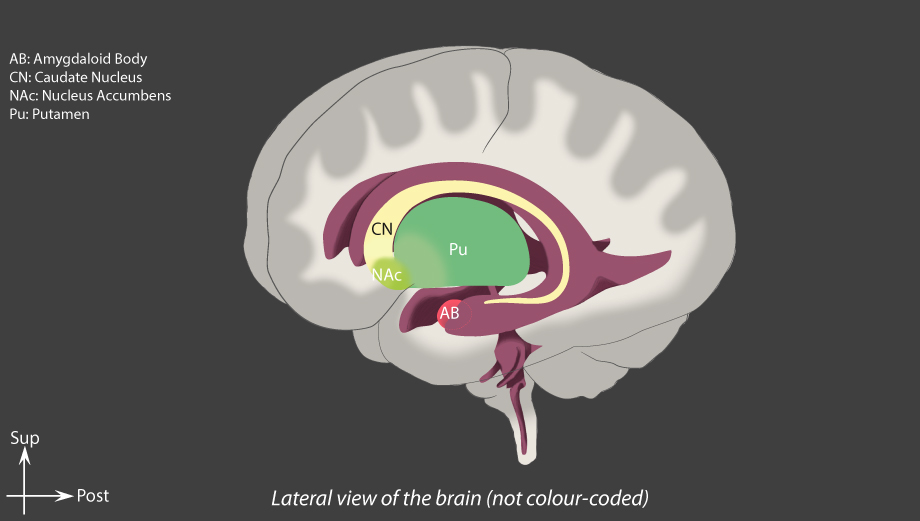
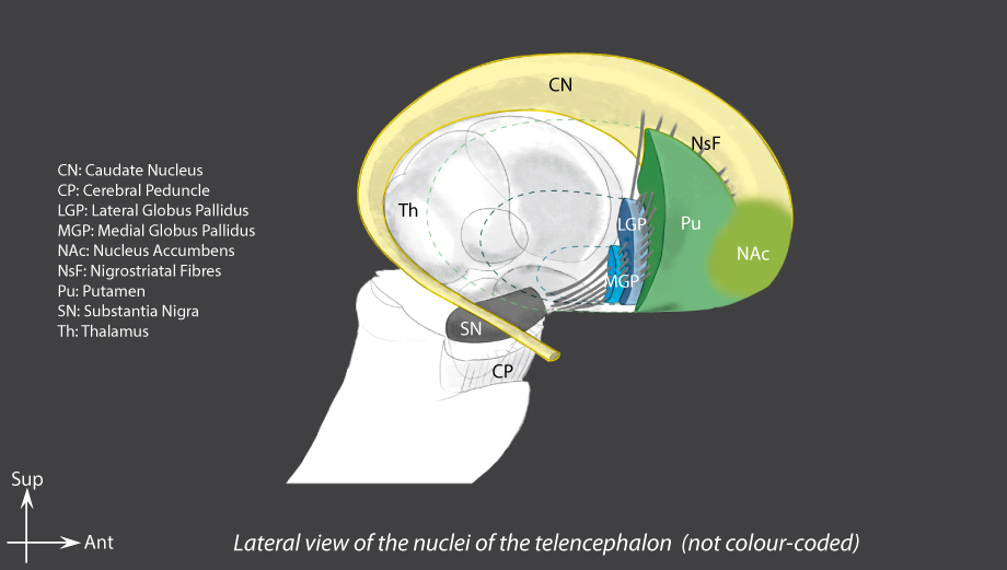
Lateral view of the brain
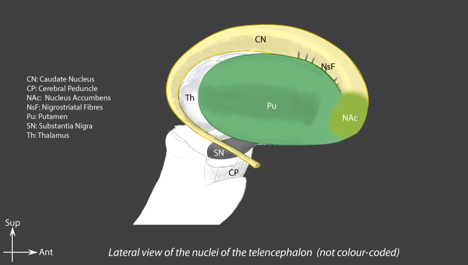
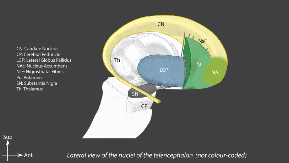
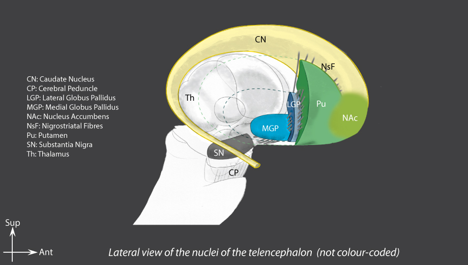
Lateral view of the nuclei of the telencephalon
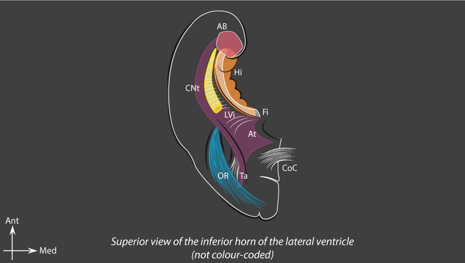
Superior view of the inferior horn of the lateral ventricles
Putamen
Front end in continuity with the head of the caudate nucleus via the nucleus accumbens
Separated from the
lateral globus pallidus
by the
lateral medullary lamina
Coronal section through the head of the caudate nucleus
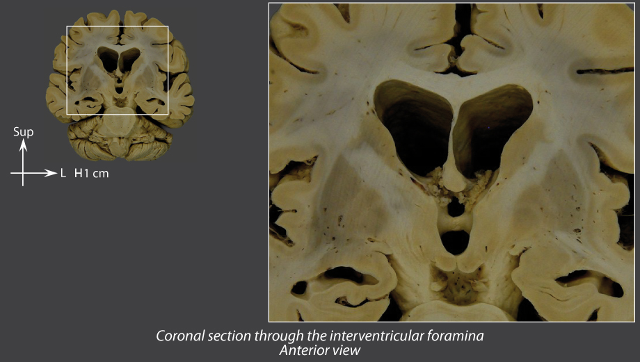
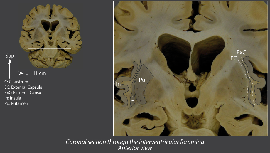
Coronal section through the interventricular foramina
Axial section through the geniculate bodies
Lateral view of the brain
Lateral view of the nuclei of the telencephalon
Globus pallidus
Divided into two parts by the
medial medullary lamina
:
Lateral globus pallidus
Medial globus pallidus
Separated from the putamen by the
lateral medullary lamina
Coronal section through the interventricular foramina
Axial section through the geniculate bodies
Innominate substance
Under the globus pallidus and the anterior commissure
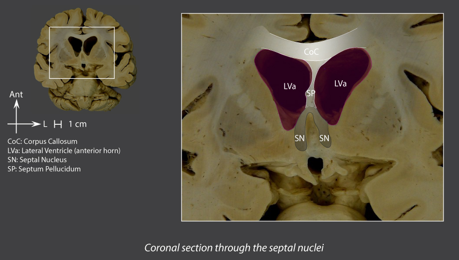
Septal nuclei
In continuity with the septum pellucidum
Claustrum
Layer of grey matter (1 or 2 mm thick)
Parallel to the inner side of the insula
Coronal section through the interventricular foramina
Axial section through the geniculate bodies
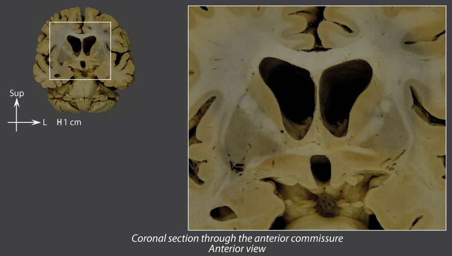
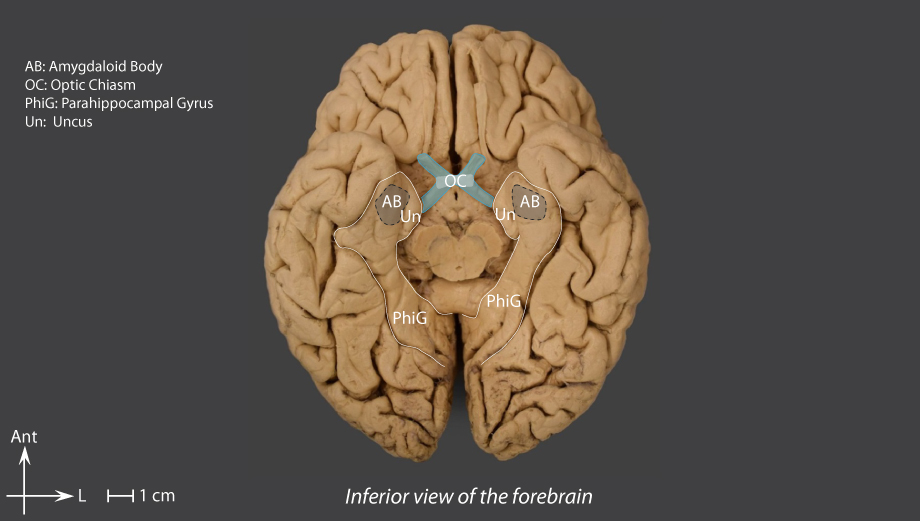
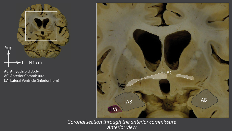
Amygdaloid body
Group of nuclei
Surrounded by the white matter of the uncus of the parahippocampal gyrus
Contributes to forming the medial front wall of the
inferior horn of the lateral ventricle
Posterior pole: extends into the stria terminalis, which runs along the inferomedial surface of the caudate nucleus to then connect to the hypothalamus
Linked to the contralateral
amygdaloid body
by the anterior commissure
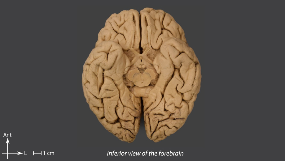
Inferior view of the forebrain
Coronal section through the anterior commissure
Superior view of the inferior horn of the lateral ventricle
Lateral view of the brain
Outline
Telencephalon
External configuration
Internal configuration – cerebral cortex
Internal configuration – hippocampus
Internal configuration – nuclei
Internal configuration – white matter
Schematic internal structure
Internal configuration – hippocampus
Internal configuration – white matter
Introduction
Telencephalon
External configuration
Internal configuration – cerebral cortex
Internal configuration – hippocampus
Internal configuration – nuclei
Internal configuration – white matter
Schematic internal structure
Diencephalon
Midbrain
Cerebellum
Pons
Medulla oblongata
Spinal cord
Meninges
Vascularization
Ventricles of the brain
Sectional anatomy
Contributors
References
Menu


 by the lateral medullary lamina