Spaces

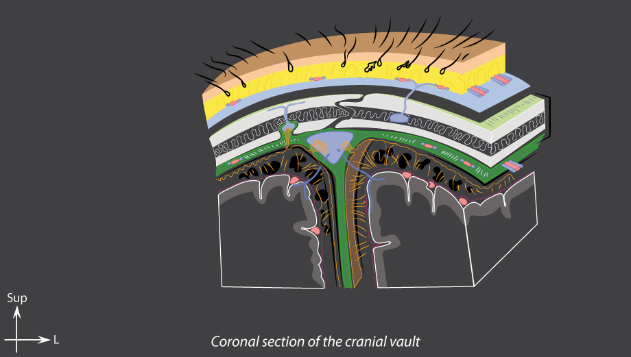
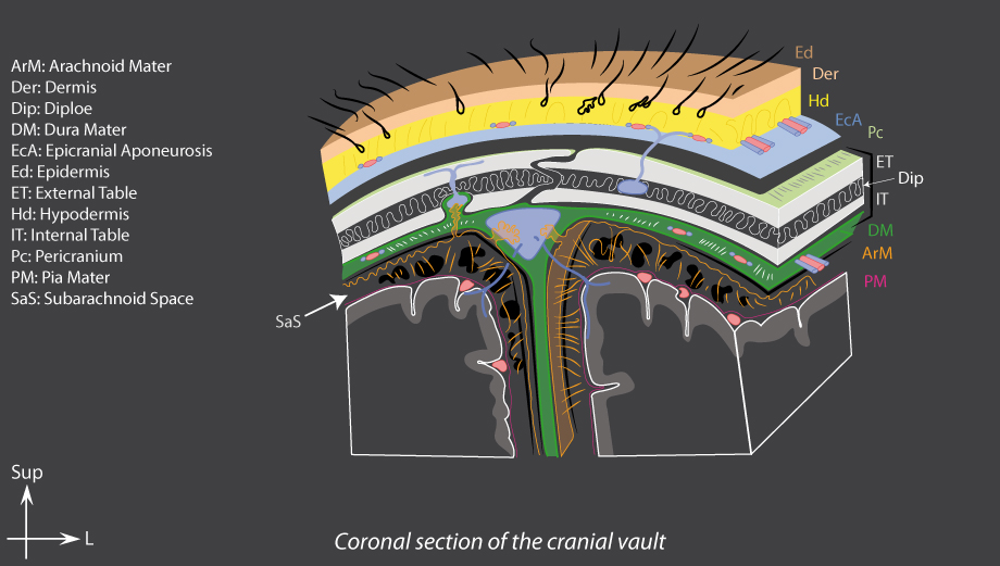
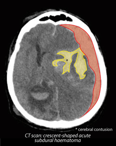
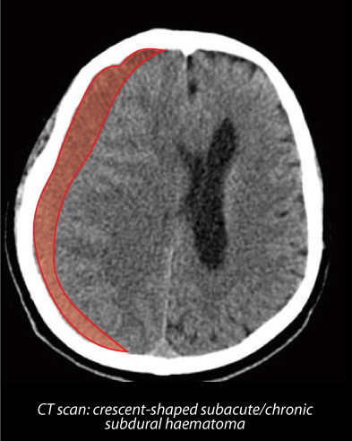
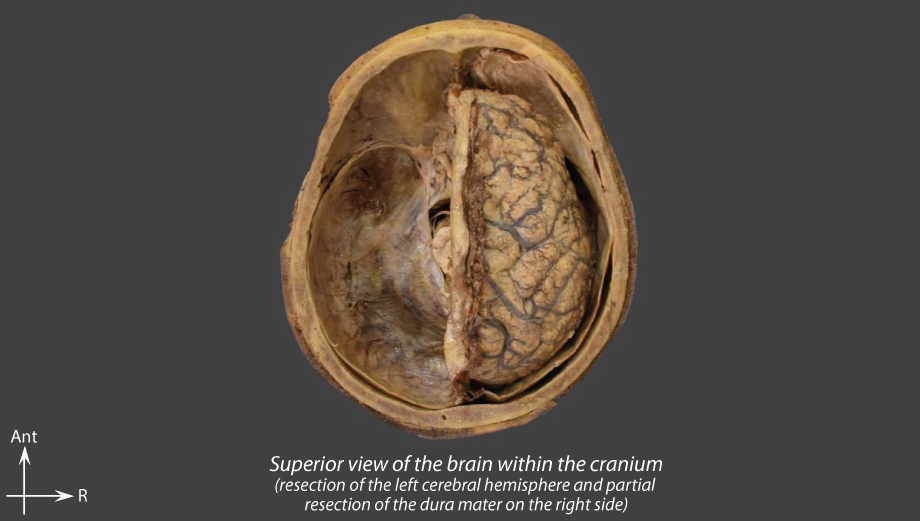
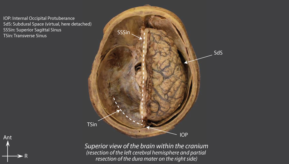
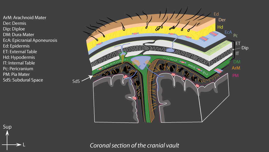
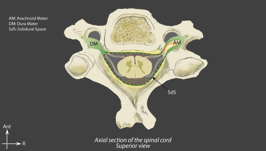
Real or virtual spaces separate the three layers of meninges:

Epidural space

Cranial
or extradural space

Extradural haematoma
  • Cause: head trauma → meningeal arteries damage
    in most cases: fracture of the temporal bone with rupture of the middle meningeal artery
  • Outcome: effusion of blood
    → the virtual epidural space becomes a real space
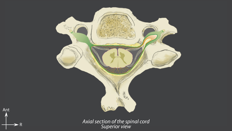
  • Clinical picture: intracranial hypertension
  • Complications: axial deviation and herniations

Spinal
ou espace péridural

  • Real space
  • Contents: epidural fat and venous plexus

Subdural space

Cranial

Subdural haematoma
  • Two types of subdural haematomas:
    Acute
    • Cause: high-velocity head trauma
    • Often accompanied by other haematomas and/or subarachnoid haemorrhaginge

    • Cause: minor head trauma, sometimes without history of trauma
    • Evolution: slow bleeding over several weeks
    • Higher incidence amongst the elderly
    • Signs of intracranial hypertension

  • Virtual space
  • Crossed by straight bridging cerebral veins draining into the venous sinuses
  • Contains CSF

Spinal

  • Virtual space

Subarachnoid space

Cranial

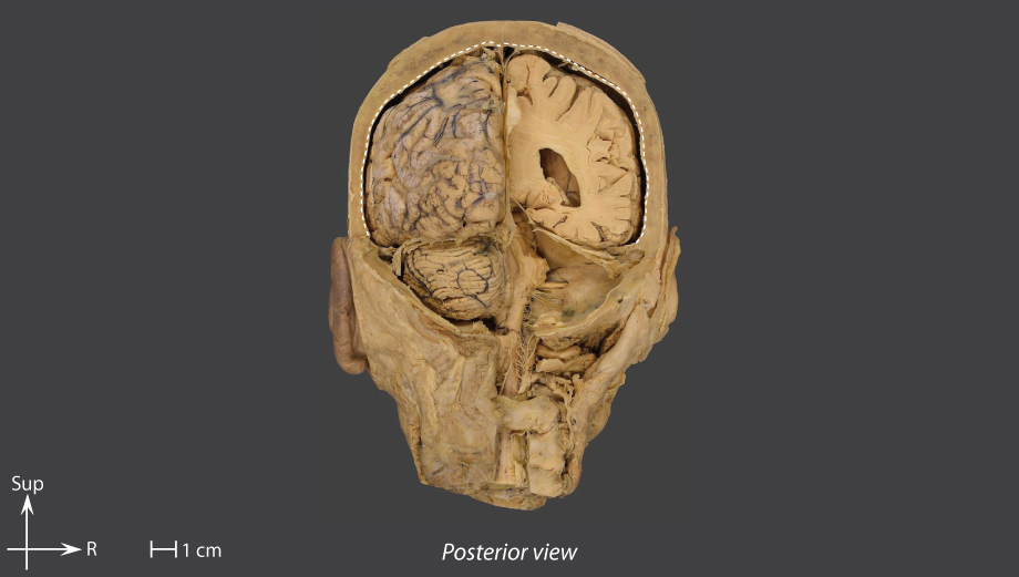
Subarachnoid haemorrhage

  • Two types of subarachnoid haemorrhage:
    Non-traumatic or spontaneous
    • Main cause: ruptured arterial aneurysm
    • Symptoms: sudden excruciating headaches
    Traumatic
    • Main cause: cerebral contusion
    • Symptoms: severe headaches

  • Real space
  • Crossed by the arachnoid trabeculae
  • Contains the cerebrospinal fluid and the large intracranial arterial vessels

Spinal

  • Real space
  • Cylindrical sheath that surrounds the spinal cord
  • Crossed by the arachnoid trabeculae
  • Contains the cerebrospinal fluid sampled by lumbar puncture: insertion of a needle in the spinal subarachnoid space, at the level of the cauda equina in order to lower the risk of injuring the spinal cord

sampled by lumbar punction: insertion of a needle in the spinal subarachnoid space at the level of the cauda equina to reduce the risk of spinal cord injury