Study of proteins adsorption on biomaterials surfaces by XPS and ToF-SIMS
Contacts : Marie
Henry, Claude Poleunis, Patrick Bertrand
The protein
adsorption is the first event happening on the surface of any system implanted
in biological environment. Once adsorbed, a protein either keeps its native
structure or undergoes changes in concentration, orientation or conformation.
These properties of adsorbed protein layers determine the subsequent cellular
interactions, which can significantly impact the performance of biomaterials in
a biological environment. The extent of the protein rearrangement is affected
by the nature and the properties of the surface. The study of the protein
adsorption is important for a lot of applications: biosensors, tissue engineering,
prosthesis, contact lenses, (bio)artificial organs, and biofouling.
Two techniques of surface analysis
are usually used to study protein adsorption on biomaterials surfaces: the
X-ray Photoelectron Spectroscopy (XPS) and the Time-of-Flight Secondary Ions
Mass Spectrometry (ToF-SIMS). XPS permits to obtain information on the adsorbed
protein amount and the thickness of the protein layer. The very high surface
sensitivity of ToF-SIMS permits to probe very low levels of protein adsorption,
which would be very interesting in studying efficiency of protein resistant
surface. In addition to allowing the identification of adsorbed proteins,
static ToF-SIMS can be used to study the conformation, the orientation and the
degree of denaturation of an adsorbed protein film. This information is
obtained in ToF-SIMS by looking at the modifications in the nature and/or in
the intensity of the amino acid SI fragments detected at the upper most surface
of the adsorbed protein. The treatment of the secondary ion peaks with
Principal Component Analysis (PCA) permits the detection of these intensity
variations.
Development
of biocompatible polymeric membranes for bioartificial pancreas
To permit the transplantation of pancreatic islets without using immunosuppressing medicines, the islets are encapsulated in an artificial membrane which permits glucose and insulin diffusion but avoids the passage of antibodies and others immunizing cells. We have two principal objectives: the development of the most appropriated membrane for the encapsulation of pancreatic islets and the comprehension of the phenomena happening at the interface between the membrane and the biological environment.
Concerning the development of the membrane, the membrane surface was studied after different surface treatments consisting mainly in covering the surface with a more hydrophilic polymer. These treatments will improve the biocompatibility and the insulin and glucose permeability.
Concerning the comprehension of the
phenomena at the interface, protein adsorption and the effect of the surface
properties on it were studied. The influence of the surface hydrophilicity on
the amount and the conformation of adsorbed human serum albumin (HSA) was
already determined. Figure 1 highlights the relation between the albumin
adsorbed amount (represented by the ratio of Ialb and Itotal)
and the albumin conformation at the surface (represented by the PC1 scores). In
the case of the native surface, there is no relation between the adsorbed
amount and the surface conformation. In the case of the hydrophilic treated
surface, there is a proportional relation between the two variables. In other
words, whatever the adsorbed amount, the albumin molecules will adopt the same
conformation on the native PC membrane. In contrast, on the treated PC
membrane, a distinct conformation of the albumin molecules corresponds to each
level of adsorbed amount.
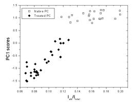
Figure 1: PC1 score plot from
PCA of positive ion spectra of HSA adsorbed on native PC membrane (open square)
or on treated PC membrane (black diamond) as a function of the ratio between
the albumin SI peak intensity and the total SI peak intensity. The PC1 scores
represent HSA conformation and the intensity ratio represents the HSA adsorbed
amount.
The adsorption of human insulin was
also studied. We have determined the ToF-SIMS fingerprint of the two proteins
(HSA and insulin). In the competitive adsorption, we can detect insulin within
the adsorbed protein layer from 5µg/ml concentration in the solution. The
membrane surface after its implantation in mini-pigs was also analysed.
This work is supported by the EC
contract BARP+ #NMP3-CT-2003-205614
Publications related to this research theme:
Henry M., Bertrand P., Surf. Interface Anal., 36 (2004) 729
M. Henry, C.
Dupont-Gillain, P. Bertrand, Langmuir 19 (2003)
6271