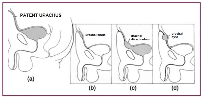
Reminder: the urachus is a median extraperitoneal tubular structure, 5-10 cm long and 5-8 mm in diameter that connects the posterior side of the umbilicus to the bladder dome and the lumen of which light is virtual. A partial or total lack of obliteration of the allantoic channel during the fifth month of gestation may be at the origin of four benign diseases of the urachus:
- an ombilico-bladder fistula (47.6 %) or omphalo-mesenteric duct. In principle, it is diagnosed in the neonatal period (a). Urine leak through the umbilicus
- an urachal cyst (30.7 %) (d): cystic mass located between the abdominal wall and the peritoneum that is repelled. The cyst does not communicate with the umbilicus, nor the bladder and is most often located on the distal third part to the urachus.
- a blind external fistula (16.4 %) or urachal sinus (b)
- a blind internal fistula (3.2 %) or urachal diverticulum (c)
Most often, the diagnosis of a pathology of the urachus is made in childhood or adolescents (62 %) after infectious or painful complications:
- urachal sinuses manifest themselves as purulent umbilical discharges or of erythematous or granulomatous periombilical lesions
- urachal diverticula vary in size, but can take massive proportions in case of association with a cervicoprostatic obstacle. They may be at the origin of a large postmicturition residue, urination in two-steps, urinary infections or complications due to urolithiasis;
- urachal cysts remain asymptomatic for a long time. They can increase in volume up to be palpable in the hypogastric region. They can also result in urinary complications, non-specific types of dysuria, hematuria and pollakiuria. Infection of urachal cysts by Staphylococcus are the most frequent. These infections can lead to peritonitis or infection of the space of Retzius.
The diagnosis is based on clinical examination and medical imaging combining ultrasound and uro-CT scan. Due to the risk of complications, surgical treatment should be systematically proposed. Omphalectomy is not recommended, but the urachus must be taken out including the lateral umbilical ligaments and a piece of bladder. There is a risk of malignant degeneration: a few cases of rhabdomyosarcoma have been reported.
Anesthetic implications:
during the neonatal period, semi-urgent surgical indication; later, urological surgery involving a risk of sepsis.
References :
- El Azzouzi D, Lasseri A.
Les anomalies congénitales de l’ouraque chez l’enfant : le point de vue du chirurgien pédiatre.
J Pédiatr & Puériculture 2013 ; 26 : 301-7.
Updated: September 2017