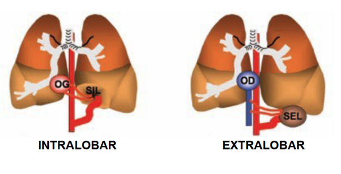
Incidence: about 1/20.000 births. Result of bronchopulmonary development abnormalities that may involve lung parenchyma, bronchi, upper digestive tract, vessels, and diaphragm. It forms a mass of non-functional lung tissue. This mass is most frequently located in the left lower lobe, in in contact with the diaphragm. The arterial vessels are systemic, usually originating from the abdominal or thoracic aorta with more than one feeder artery in 15 % of cases. Venous drainage is variable, into the pulmonary veins, the inferior vena cava or the azygos system.
Intralobary and extralobar sequestrations are distinguished according to the presence or absence of visceral pleura and their venous drainage.
- Extralobar forms:
1) they are often associated with other malformations: diaphragmatic hernia, for example,
2) they may be localized in the mediastinum or in the abdominal cavity (subdiaphragmatic form).
3) they are covered with a specific visceral pleura
4) they are vascularized by an abnormal branch originating from the thoracic or abdominal aorta; rarely, this feeding artery can come from a subclavian artery, coronary artery or the celiac trunk.
5) they have a systemic venous drainage: azygos vein in general, but cases of hemiazygos drainage, inferior caval or portal vein have been described.
6) preoperative angiography is therefore essential. Bilateral forms are described that can lead to neonatal cardiac failure.
- Intralobar sequestration
1) they are rarely congenital: they are usually the result of pneumonitis: local inflammation leads to the recruitment of an abnormal systemic artery to vascularize an initially healthy lung segment. The pulmonary segment contains dense sometimes cystic parenchyma
2) they are covered by the same visceral pleura as the normal lung
3) they drain into the pulmonary venous system
4) they are often silent and discovered by hazard; but they can result in repeated infections that should one suspect a bronchial connection. Their radiological appearance depends on the degree of aeration of the lesion and the possible existence of an associated lesion.

Some cases, called hybrids, associate pulmonary sequestration and airway malformation (see this term).
Clinical presentation:
- prenatal screening (diagnosis during the second trimester of gestation). The more or less echogenic lesion, connected to the aorta by a vessel (Doppler ultrasound), generally grows until the 26th week and then stabilizes or regresses, or even disappears. It can also lead to hydramnios or hydrops foetalis and require in utero treatment: corticosteroids, amniotic shunt ... If the child is asymptomatic at birth and does not have a mediastinal shift, treatment consists of regular monitoring of the size of the lesion.
- fortuitous discovery following frequent respiratory infections, feeding difficulties,
- signs of cardiac failure secondary to a left-right shunt or arteriovenous fistula.
The treatment is surgical, by thoracotomy or thoracoscopy.
Pulmonary sequestrations are related to other rare malformations, such as:
- Scimitar syndrome (see this term), that almost exclusively affects the right lung where the lower lobe is drained at the subdiaphragmatic level into the inferior vena cava or a hepatic vein; usually other malformations are associated such as a right pulmonary hypoplasia and/or a partial vascularization by an aberrant systemic artery; chest X-Ray shows dextroposition of the heart and a scimitar aspect of the abnormal vein
- systemic arterial vascularization of a lung lobe, generally the lower lobe; venous drainage is normal through the pulmonary veins;
- accessory diaphragm syndrome: the hemithorax, most often the right one, is divided into 2 compartments by a membrane derived from the pericardium; it combines a homolateral pulmonary hypoplasia with a partial anomalous venous return in the inferior vena cava (frequent), an abnormal systemic vascularization and, more rarely, a diaphragmatic hernia as well as vertebral anomalies;
- horseshoe lung: malformation where the lungs are fused posteriorly or where one of the lungs is hypoplastic; it is associated with dextrocardia, cardiac malformation (ASD, VSD, double outlet ventricle) and often, as in the accessory diaphragm syndrome, to a partial abnormal venous return in the inferior vena cava as well as an abnormal systemic vascularization. Important risk of hypoxemia and of pulmonary hypertension.
Anesthetic implications:
check the hemoglobi nsaturation at room air; evaluation of the systemic vascularization and the venous drainage of the lesion; Sometimes preoperative embolization of the feeding artery is necessary before considering surgical resection; significant hemorrhagic risk; decide with the surgeon if unipulmonary ventilation could be useful. Thoracic epidural or paravertebral block for postoperative analgesia.
References :
- Mendeloff EN.
Sequestrations, congenital cystic adenomatoid malformations, and congenital lobar emphysema.
Semin Thorac Cardiovasc Surg 2004; 16:209-14.
- Von Scheidt F, Eicken A, Wowra T, Brunner H, Apitz C.
Bilateral pulmonary sequestration in a preterm infant.
J Pediatr 2018; 194: 260-1.
- Sato A, Morita M, Ota H, Kamimura Y, Ito H, So M Tamura T et al.
General anesthetic management of a child with horseshoe lung and left lung hypoplasia for cheiloplasty: a case report.
A&A Practice 2018; 11: 208-12.
- Leblanc C, Baron M, Desselas E, Hanh Phan, Rybak, Thouvenin G1, Lauby C, Irtan S.
Congenital pulmonary airway malformations: state-of-the-art review for pediatrician’s use.
Eur J Pediatr 2017 ; 176:1559-71
- Johansen M., Veyckemans F., Engelhardt T.
Congenital anomalies of the large intrathoracic airways.
Pediatr Anesth 2022; 32: 126-37
- Karlsson M, Conner P, Ehren H, Bitkover C, Mesas Burgos C.
The natural history of prenatally diagnosed congenital pulmonary airway malformations and bronchopulmonary sequestrations.
J Pediatr Surg 2022 ; 57 : 282-7
Updated: November 2022