Rare. It is a blown cavity filled with air, developed in the lung parenchyma.
The main causes are:
- a sequel of acute pneumonia, including to S aureus, P aerogenisus or M pneumoniae : it is a consequence of the drainage of parenchymal necrosis with creation of a valve system by inflammatory exudate at the bronchiolar level
- chest trauma: a bronchiolar rupture occurs during a brutal decompression of the chest following a violent shock.
- barotrauma: secondary to an alveolar excess pressure in a closed glottis trauma.
The clinical symptomatology is poor, mainly dominated by dyspnea or hemoptysis in case of traumatic cause.
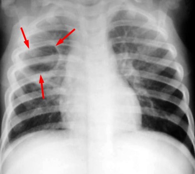
Classical radiological aspect is that clarity of regular outlines, sometimes multifoil, often unique. Its size is variable, sometimes exceeding 10 cm in diameter. The wall is thin, almost invisible, sometimes thicker when it is surrounded by a parenchymatous condensation. These lesions are often masked by the adjacent parenchymatous lesions. The chest CT scan is more sensitive to evaluate the lesion. The evolution is favourable in the majority of cases without sequelae with the pneumatocele regression with an average delay of 2 weeks. Complications are rare: superinfection, healthy parenchyma compression or even of the mediastinum, rupture into the pleura leading to a pneumothorax. Percutaneous drainage of the pneumatocele is often carried out under RX guidance .
Differential diagnosis: an intra-parenchymatous bronchogenic cyst, bubbles of emphysema, blebs, hydatic cyst
completely evacuated by a vomica in an endemic area, lung abscess.
Anesthetic implications:
keep that possibility in mind if recent history of pneumonia or empyema; induction in spontaneous breathing (to avoid dilating the cavity by positive pressure) and selective intubation of healthy lung.
References :
- de Bie HMA, van Toledo-Eppinga L, Verbeke JIML, van Elburg RM.
Neonatal pneumatocele as a complication of nasal continuous positive airway pressure.
Arch Dis Child, Fetal & Neonatal Ed, 2002; 86: F202-3 - Liu C-M, Hang C-H, Lau H-P, Yeh H-M.
Anesthetic management for two infants undergoing surgery for tension pneumatoceles.
Pediatr Anesth 20017; 17: 189-90
Updated: March 2016