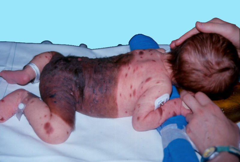
Rare dysembryoplasia (1/20,000 to 1/500,000 births) that seems more common in girls and in the Asian population. It is the consequence of incomplete and abnormal migration of the nevoblasts before the end of the 3rd month of gestation. The nevus is present from birth and it is histologically a nevocellular or mixed nevus, rich in melanin and with a very important vascular development. Its size is variable: it is called giant when its maximum diameter is greater than 20 cm.
Clinically, it is a large dark thick skin area with black spots, the borders of which are sharp but irregular. Moreover, its surface is irregular (nodules of proliferation), sometimes warty or hairy. Its location is usually mediodorsal, often symmetrical ('as a cape'), or as 'underpants', if it covers the urogenital area. More or less large isolated nodules can be found on other parts of the body.
In case of involvement of the neck or face, neuromeningeal melanosis (with a risk of epilepsy) is often associated.
Two problems:
- risk of malignant degeneration: about 5 %, which is 16 x higher than in the general population; these melanomas occur frequently before the age of 7 years.
- esthetic and social aspect.
Different surgical techniques can be used: excision/ suture (small surfaces): for large surfaces, advantage is taken of a cleavage plane between the superficial and deep dermis to achieve 'surface' interventions: curettage, dermabrasion, laser, chemical peeling, hydrosurgery ('Karcher') ; later: excision-graft.
Hypophosphatemic rickets may be associated. The association of hypophosphatemic rickets with skin manifestations of epidermal nevus or pigmento-keratotic phacomatosis defines the skeletal and cutaneous hypophosphatemic syndrome. It is due to identical somatic activating RAS mutations, identical in the skin and bone, (NRAS, KRAS and HRAS). Brain stem lipomas associated with epidermal nevi have been reported. The treatment of this rickets consists in giving 1-alpha and supplements of phosphates.
Anesthetic implications:
(1) neonatal anesthesia with risk of hypothermia; (2) painful surgery with important fluid and/or blood losses (depending on the surgical technique): post-operative care similar to those of a burned patient if the abraded surface is large: ICU admission is therefore recommended. (3) during the post-operative period, baths and dressings under anesthesia. (4) sometimes important pruritus (source of agitation and distress) during skin regrowth 5) multiple surgeries
References:
- Arneja JS, Gosain AK.
Giant congenital melanocytic nevi.
Plast Reconstr Surg 2007 ; 120 : e26-40.
- Makkar HS, Fieden IJ.
Congenital melanocytic nevi: an update for the paediatrician.
Curr Opin Pediatr 2002; 14: 397-403.
- Marghoob AA, Borrego JP, Halpern AC.
Congenital melanocytic nevi: treatment modalities and management options.
Semin Cutan Med Surg 2007; 26: 231-40.
- Warner PM, Yakuboff KP, Kagan RJ, Boyce S,Warden GD.
An 18-year experience in the management of congenital melanocytic nevi.
Ann Plast Surg 2008; 60: 283-7.
- Welfringer-Morin A,
Pinto
G, Baujat G,
Cavé
H, de Saint Denis
T, Hadj-Rabia
S, Bodemer
C, Boccara O.
Rachitisme hypophosphatémique : complication rare du syndrome du naevus pigmentaire congénital.
Ann Dermatologie Vénéréologie ; 2018 :145, S20
Updated: November 2021