Type 1:
Incidence: 1/10,000. Systemic connective tissue disease most often due to a mutation in the fibrillin 1 gene (FBN1) on 15q21.1. Autosomal dominant transmission but sporadic in 25 % of cases . Some cases are due to a mutation in the TGFBR2 gene (chr 3) that codes for a receptor of TGF-beta (see Loeys-Dietz syndrome).
Association to varying degrees of:
- arachnodactyly: very long fingers
- dolichostenomelia: long arms and legs
- joint hyperlaxity with frequent dislocations
- micrognathia, arched palate, malar hypoplasia
- dislocation of the lens, megalocornea, myopia, retinal detachment (more rarely: glaucoma, cataract)
- pectus excavatum or carinatum
- progressive dilation of the aortic arch with risk of rupture at the end of adolescence; this expansion also causes a progressive aortic insufficiency.
- mitral valve prolapse or true mitral insufficiency (with risk of arrhythmias, endocarditis, congestive heart failure)
- scoliosis
- dural ectasies (65-90 % of cases, especially at the lombosacral level): pulsatile pressure exerted by the cerebrospinal fluid on the fragile dura mater leads to its gradual expansion, with an enlargement of the dural sac in young adults and, gradually, of the spinal canal, especially at the lombosacral level. They can cause lombosacral or genital pain, or even spontaneous leptomeningeal breachs with leak of CSF following an effort (orthostatic headache).
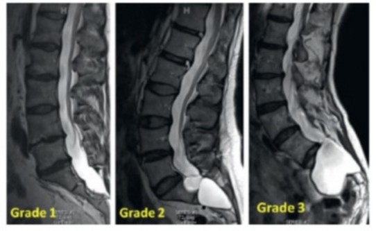
Fattori's classification:
- grade I: ectasia of the spinal canal and disappearance of the epidural fat at the level of one vertebral body
- grade II: bulging of the dural sac with erosion of the posterior wall ('scalloping') of some vertebral bodies
- grade III: sacral meningocele
Type 2:
also called Beals syndrome (see).
Medical treatment: β-blockers decreases the rate of dilation of the ascending aorta; it seems that a treatment with angiotensin receptor inhibitors is more effective
Anesthetic implications:
careful positioning (risk of dislocations). Increased risk of pneumothorax. Cardiac ultrasound (aortic and mitral valves). Critical importance of maintaining hemodynamic stability.
In case of dural ectasies (young adult), spinal anesthesia may be less effective (dilution of the local anesthetics) and it is better to perform an epidural block, in which case the dose can be more easily titrated. It is unclear if there is a higher risk of dural tap. It is wise to obtain an imaging of the sacral canal (ultrasound ?) before performing a caudal block.
In case of pregnancy, the evolution of aortic dilatation appears to be accelerated; the current recommendations are:
- measurement of the diameter of the aortic arch before pregnancy: avoid any pregnancy if it is > 45 mm; discuss case by case if it is between 40 and 45 mm
- treatment with ▀-blockers
- quarterly monitoring of the diameter of the aorta (and every month in the 3rd quarter of pregnancy)
- if the diameter of the aortic arch is < 40 mmĀ: delivery by natural route with epidural analgesia (avoiding efforts to push: forceps or vacuum extraction)
- if the diameter of the aortic arch is > 45 mm, the aortic has already been replaced or the diameter of the aortic arch has highly increased during pregnancyĀ: elective caesarean section around 36 weeks of gestation
- the presence of a scoliosis (operated or not) renders the achievement of an neuraxial block difficult
- the goal of general anesthesia for caesarean section is to avoid hypertensive peaks: invasive monitoring of BP, administration of an opioid before intubation (sufentanil, remifentanil ...)
References :
- Morse RP, Rockenmaster S, Pyeritz RE, Sanders SP, Bieber FR, Lin A, McLeod P, Hall B, Graham JM.
Diagnosis and management of infantile Marfan syndrome.
Pediatrics 1990; 86: 888-95. - Oh AT, Kim YH, Kim BK, K H-S, Kim CS.
Unexpected tracheomalacia in Marfan syndrome during general anesthesia for correction of scoliosis.
Anesth Analg 2002; 95: 331-2. - Mokri B.
Low cerebrospinal fluid pressure syndromes.
In Neurol Clin N Am. Elsevier-Saunders 2004; 22: 55-74. - Baghirzada L, Krings T, Carcalho JCA.
Regional anesthesia in Marfan syndrome, not all dural ectasias are the same: a report of two cases.
Can J Anesth 2012; 59: 1052-7. - Lacassie HJ, Millar S, Leithe LG, Muir HA, Montana R, Poblete A, Habib AS.
Dural ectasia: a likely cause of inadequate spinal anaesthesia in two parturients with Marfan’s syndrome.
Br J Anaesth 2005; 94: 500-4. - Leyla Baghirzada, Timo Krings, Jose C.A. Carvalho
Regional anesthesia in Marfan syndrome, not all dural ectasias are the same: a report of two cases.
Can J Anesth 2012; 59:1052-7 - Allyn J, Guglielminotti J, Omnes S, Guezouli L, Egan M, Jondeau G, Longrois D, Montravers P.
Marfan’s sydrome durig pregnancy : anesthetic management of delivery in 16 consecutive patients.
Anesth Analg 2013; 116: 392-8. - Sakurai A, Miwa T, Miyamoto Y, Mizuno Y, Ka K.
Inadequate spinal anesthesia in a patient with Marfan syndrome and dural ectasia.
A&A Case Reports 2014 ; 2 : 17-9 - Ackerman I, Henze G, Kottke R, Weiss M.
Spinal meningeal cyst in a child with Marfan syndromeĀ: a potential cause for apparent dural puncture during caudal epidural block.
Pediatr Anesth 2019; 29: 959-61. - Berger JA, Huh DD, Lee T, Wadia RS, Bembea MM, Goswani DK.
Perioperative management and considerations in pediatric patients with connective tissue disorders undergoing cardiac surgery.
Pediatr Anesth 2021Ā; 31Ā: 820-6.
Updated: February 2022