Rare: 0.5 to 1 /106/year. Most common pediatric bullous disease. Autoimmune origin. It is possible that the HLA Cw7 group be genetically more susceptible.
Two forms:
- primary form: in childhood (from 1 to 12 years of age, peak around 5 years of age), sometimes associated with an IBD (Inflammatory Bowel Disease)
- secondary form: after puberty or adulthood, often secondary to a medication (2 to 30 days interval between the medication intake and the onset of lesions): vancomycin, amiodarone, NSAID's, penicillins, antiepileptics. Rapid healing when drug intake is stopped
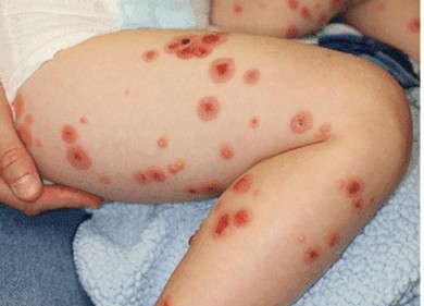
Clinical presentation: skin or mucous pruriginous blisters, often organized in a rounded or arciform way (rings); generally located at the lower part of the trunk (periombilic), on the limbs and the perineum; mucous membrane involvement is possible (mouth, conjunctiva)
Diagnosis: biopsy of a lesion showing a picture of epidermal vesicle with micro abscess of polynuclear neutrophils) and above all, at immunofluorescence, a linear and homogeneous deposition of IgA along the basal membrane.
Treatment:
- diaminodiphenylsulfone (Dapsone®): 0.5 to 2 mg/kg/d
- in case of failure or contraindication: sulfapyridine 1 to 3g/day or colchicin 0.5 to 1 mg/kg/day.
- sometimes dermocorticoids or systemic corticosteroids
Anesthetic implications:
check total blood count and liver function in case of treatment with Dapsone® or colchicin. Risk of methemoglobinemia in case of Dapsone® treatment; check for oral lesions
References:
- Costenoble A, Tennstedt D.
Dermatose bulleuse ŕ IgA linéaire.
Louvain Médical 2018; 137: 203-8
Updated: March 2025