Unknown prevalence. In adults, women and men are equally affected, but in young children, the sex ratio could be 1.5 boys for 1 girl. It is the most common form of non-Langerhansian histiocytosis, one of the 3 classes of histiocytic diseases. Histiocytic diseases are reactive or malignant diseases in which various tissues, including the skin, are infiltrated by cells of the monocyte-macrophage lineage.
A particular form of xanthogranulomatosis is ALK-positive Histiocytosis (Anaplastic Lymphoma Kinase) linked to a translocation of the ALK gene at 2p23, and classified into 3 forms:
* 1A: systemic form: liver, spleen, hematopoietic system
* 1B: systemic form: central nervous system, lung, bone, skin, lymph nodes
* 2: monosystemic form: skin or central nervous system
Hypothesis: the histiocytosis of the dermal dendrocytes or dendritic cells could constitute a spectrum of diseases where the dermal dendrocyte is present at a different stages of maturation. In case of xanthogranulomatosis, these cells are of the "young" (immature) type and associated with brief diseases, healing spontaneously within a few months.
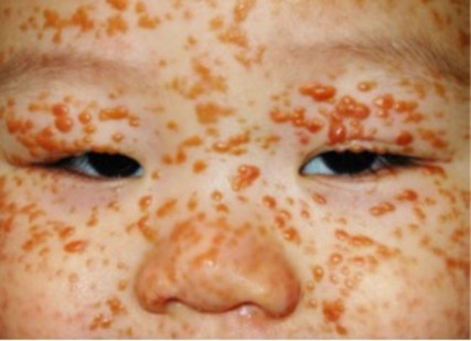
Juvenile xanthogranuloma most often appears on the skin of the baby or young child as nodules composed of histiocytic cells, but it sometimes develops in the eyes or viscera. It is unique in more than 60 % of cases. Xanthogranulomas are usually asymptomatic but may (rarely) ulcerate or bleed. The lesion is usually benign and progresses to self-healing, but can be problematic when it develops in the eye (0.3 to 0.5 %) (risk of glaucoma or hyphema) or viscera.
Other possible extracutaneous manifestations are in order of decreasing frequency:
- pulmonary: dyspnea, rounded opacities resembling metastases.
- hepatic: hepatomegaly.
An adult xanthogranuloma form has been described : it is benign and heals spontaneously but very rarely presents necrobiotic forms.
Forms with multiple nodules may be associated with neurofibromatosis type 1: in these cases, the risk of subsequent myelomonocytic leukemia is greatly increased.
Clinical presentation :
¨ external appearance: clearly delineated, smooth and firm (rubber consistency) round to ovale papules. Their color, pink to red, is tinged with yellow and tends to turn yellow-brown over time
¨ these papules appear generally on the head and neck, less often on the upper trunk and limbs and very exceptionally on the oral mucous membranes. They are sometimes hyperkeratotic or taking giant shapes, or pedunculated.
¨ size is variable; from 5 millimeters in diameter and a few mm thick to a clearly visible tumor several centimeters wide (up to 2.5 cm , with some rare cases of giant shape (macronodular, up to 10 cm can be confused with a hemangioma)
¨ xanthogranuloma is more often isolated (60 % to 82 % of cases), and present on the head (possibly on the scalp), neck, trunk and limbs. There are two variants that often coexist:
- nodular shape small, with many hemispheric papules of 2-5 mm in diameter,
- nodular form with only one or a few larger (10 to 20 mm in diameter) nodules.
Histology: xanthogranulomas initially contain non-lipid histiocytes, without Langerhans cells. Later inflammatory cells (including foamy histiocytes) appear, progressively more numerous with the maturation of the tumor. Giant cells of Touton, with multiple nuclei organized in a ring, are typical.
Treatment: in systemic forms (5 % of cases, often associated with an ALK gene translocation at 2p23), the treatment is similar to Langerhans histiocytosis: prednisolone, vinblastine and cladribine. Good results have been obtained with oral alectinib, an ALK (anaplastic lymphoma kinase) inhibitor.
.jpg)
Anesthetic implications:
none in case of an isolated form. Risk of glaucoma in case of ocular involvement. Check blood count if combined with neurofibromatosis type 1.
References :
- Kupfer-Bessagueta I, Starozb F, Plantina P.
Xanthogranulome juvénile.
Ann Dermatol Vénéréol 2009 ; 136, 70-3
- Chen T, Ma D-L.
Multiple Juvenile Xanthogranulomas.
J Pediatr 2022;251:210-1.
- Xu J, Yao X, Wen Y, Lian H, Han X, Xu Z.
Alectinib in the treatment of systemic Juvenile Xanthogranuloma of infancy with ALK translocation.
JAMA Dermatology 2023 ; 159 :1399-1401
- Wu P-C, Chen K-Y.
An infant with progressive yellowish papules and nodules.
JAMA Dermatology 2024 ; 160 : 775-6
Updated: July 2024