This is the presence of glial tissue (brain support cells) outside its normal location, i.e. outside the central nervous system. The most common site is the nose, but other ENT sites have been described: oral cavity, ear, tonsils, orbit. One case of pulmonary localization has been published.
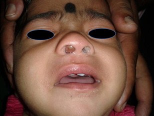
Nasal glial heterotopia usually appears at birth or in early childhood (rarely in adulthood). It is a non-pulsatile mass that can lead to nasal obstruction, deformation of the nasal septum and nasal bone, and respiratory distress if left untreated. Although nasal glial heterotopia does not communicate with the central nervous system, a cribriform plate abnormality is sometimes associated.
Histologically: mature astrocytes and glial cells in a fibrovascular stroma. Ependymal elements are often present.
Differential diagnosis: other ENT tumors, including lingual thyroid, lymphatic malformation, thyroglossal cyst or teratoma. Pulmonary malformation in case of pulmonary localization.
.jpg)
Anesthetic implications:
depending on tumor localization; risk of bleeding.
References :
- Tournier L, Berrebi D, Peuchmaur M, Bonnard A, Belarbi N, et al..
Hétérotopie gliale pulmonaire : une lésion exceptionnelle chez un nourrisson avec un jumeau anencéphale.
Annales Pathol 2019; 39: 24 -8.
- Gallego Compte M, Menter T, Guertler N, Negoias S.
Nasal glial heterotopia: a systematic review of the literature and case report.
Acta Otorhinolaryngol Ital 2022 ;42 :317-24.
- Quatrea R, Baguanta A, Gila H, Schmerber S.
Glial heterotopia of the middle ear.
European Annals of Otorhinolaryngology, Head and Neck diseases 2020 ; 137 : 207-9
- Bonaventure CA, Stark MW, D’Souza JN.
Oral mass in an infant.
JAMA Otolaryngology–Head & Neck Surgery online May 29, 2025
Updated: June 2025