(Monostotic fibrous dysplasia, polyostotic fibrous dysplasia)
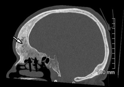
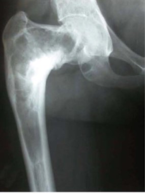
Accounts for about 2.5 % of bone tumors. Congenital bone disease where normal bone is replaced by more fragile pseudofibrous tissue following a somatic mutation of the GNAS gene (20q13.32). Lesions usually appear in childhood. All bones can be affected, but the most frequently ones are the ribs (45 %), the femur and the bones of the skull. The main symptoms are pain, deformities and a greater risk of fracture. The diagnosis is confirmed by X-ray.
The polyostotic form (20 to 30 % of cases) is often more severe and mainly affects the femur (90 % of cases), the tibia (80 %), the pelvis (80 %), as well as the ribs, the bones of the skull and face, arms and vertebrae. The lesions are usually lateralized on the same side: they are diagnosed before the age of 10 years in 2/3 of cases.
Special forms:
- the polyostotic form can be associated with skin and endocrine abnormalities: this association results in the McCune Albright syndrome (see this term).
- the association with intramuscular myxomas is known as Mazabraud syndrome. Myxomas, often multiple, are close to bone lesions. Their excision is often followed by a recurrence, but a sarcomatous degeneration is exceptional.
- hypophosphatemic osteomalacia due to tubular loss of phosphates (or phosphate diabetes) is present in about one in 2 cases.
Treatment: sometimes IV biphosphonate. In case of hypophosphatemia, it is necessary to normalize phosphatemia, by administering phosphorus supplements by mouth, systematically associated with vitamin D3. This treatment leads to a risk of hypercalciuria and therefore urolithiasis or nephrocalcinosis.
Anesthetic implications:
risk of pathological fracture; in case of polyostotic form, check phosphatemia and renal function
References :
-
Updated: May 2022