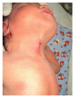
Very rare. Congenital anomaly located on the anterior midline of the neck and resulting from a defect of mesodermal fusion of the two first branchial arches.
Clinical presentation: red looking and atrophic skin path, located at the anterior part of the neck and on the midline. It is often associated to a subcutaneous fibrous cord, and the presence of a fistulous opening remaining at its caudal end as well as a structure of mamelonary aspect at its upper part. Sometimes a cyst is included. This malformation can extend from the submental region to the sternal notch, but does not, on average, exceed one third of this distance.In general, it is an isolated malformation. In case of extension to the mandible, lower lip, tongue or the sternum, it is important to look for associated anomalies, including cardiac ones.
Anesthetic implications:
difficult intubation in the presence of micrognathia or an extensive subcutaneous fibrous cord.
References :
- King J, Patel RV, Huddart SN.
Congenital midline cervical cleft.
J Pediatr Surg Case Reports 2013; 1: 99-101
- Goldfisher R, Bawa P, Ibrahim Z, Amodio J.
Clinical and imaging features of a congenital midline cervical cleft in a neonate: a rare anomaly.
Case Reports in Pediatrics 2015; doi 10.1155/2015/439596
- Achard S, Leroy X, Fayoux P.
Congenital midline cervical cleft: a retrospective case series of 8 children.
Int J Otorhinolaryngol 2016; 81:60-4
Updated: February 2021