Prevalence: about 1/5000 births. Defect of closure of the cephalic part of the neural tube (4th week of gestation) which causes the protrusion of meninges (meningocele or encephalocele, accompanied by brain tissue) at the level of a portion of the skull.
The most frequent locations are:
- the occiput (75 %): the defect is located on the midline in 90 % of cases
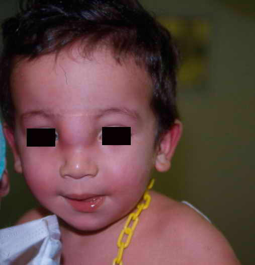
- the fronto-ethmoidal region ( syncipital encephalocele): nasofrontal, nasoethmoidal, nasoorbital (figure)
- the basal region (1-10 %): transethmoidal: nasal obstruction (sometimes hypertelorism, cleft lip and palate, bifid nose)
- the parietal region
The encephaloceles are associated with other neurological abnormalities in more than 60 % of the cases:
- hydrocephalus, microcephaly
- developmental disorders
- convulsions
- spina bifida
- Dandy-Walker syndrome
- Arnold-Chiari type II malformation.
Ocular abnormalities are frequent in case of encephalocele in the nasal region.
Except in case of leakage of CSF or respiratory problems, the surgical closure is performed as an elective procedure. The most frequent postoperative complications are hydrocephalus and meningitis.
The cure of an occipital encephalocele is performed in the prone position.
Some teams correct nasal encephaloceles endoscopically (endonasal approach).
nasal encephalocele
Anesthetic implications:
- have the contents of the mass (nerve tissue? vessels?) checked (MRI, ultrasound) before planning surgery
- in the case of a mass in the fronto-ethmoid region: check pituitary function (thyroid, growth hormone, ADH, etc.)
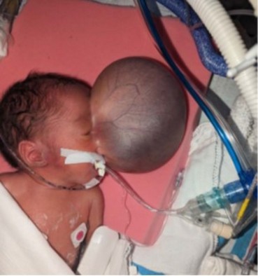
- anticipate difficulties with 1) face mask ventilation in case of a nasal encephalocele; 2) intubation if the mass is in front of the nostrils (figure 2) or in the transpalatal region; 3) positioning for induction and intubation (lateral decubitus or head held at the end of the table by an assistant, partial decompression in the case of a giant liquid mass) in the case of occipital encephalocele.
- caution: compression of the mass may cause acute intracranial hypertension. Significant hemodynamic changes may occur during incision (abrupt loss of a large volume of CSF and interaction with the brain stem) and when the herniated part of the brain is reintegrated into the skull (intracranial hypertension).
References :
- Leelnukrom R, Wacharasint P, Kaewanuchit A.
Perioperatve management for surgical correction of frontethmoidal cephalomeningocele in children: a review of 102 cases.
Pediatr Anesth 2007; 17: 856-62.
- Arora S, Bharti N, Bala I.
Sudden cardiac arrest during repair of giant occipital encephalocele with microcephaly.
J Anaesth Clin Pharmacol 2010; 26: 259-60.
- Mahajan C, Rath GP, Dash HH, Bithal PK.
Perioperative management of children with encephalocele: an institutional experience.
J Neurosurg Anesthesiol 2011; 23: 352-6.
- Vasudevan A, Kundra P, Priya G, Nagalakshmi P.
Giant occipital encephalocele : a new paradigm.
Pediatr Anesth 2012 ; 22 : 586-8
- Black SA, Galvez JA, Rehman MA, Schwartz AJ.
Images in Anesthesiology : airway management in an infant with a giant occipital encephalocele.
Anesthesiology 204: 120:1504
- Nhushan RV, Pratibha CB, Balasubramanya AM, Nandakumar R, Reunold R.
Endoscopic management of nasal encephalocele in a 4-month-old baby.
Int J Pediatr Otorhinolaryngol Extra 2014; 9: 67-9.
- Fandino A, Herrera AJ, Feo OE, Ronderos AM, Ospina JC, De Leon O.
Congenital meningocele associated with corpus callosum agenesis and midline superior labial cleft due to Tessier 0-14 facial fissureĀ: a case report.
Int J Pediatr Otorhinolaryngol 2020; 130: 129Ā:109764
- Vogler A, Tovar A, Kim E, Le S, Lo C.
A giant basal meningoencephalocele repair in a neonate: a case report.
A&A Practice. 2024;18:e01821
- Ma R, Brown J, Careskey M .
Perioperative management of massive anterior encephalocele in a newborn: a case report.
A&A Practice. 2024;18:e01809
Updated: August 2024