Incidence: around 1/6000 births, with a male preponderance: 1.4 boys to 1 girl. Familial forms are rare. It is the result on an abnormality in the early development of the primitive intestine.
It may be :
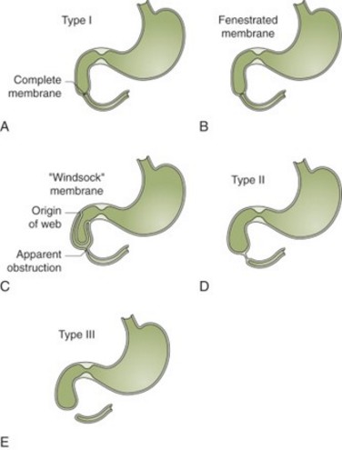
- complete: atresia: high occlusive syndrome with bilious vomiting due to the presence of:
- a diaphragm in the intestinal lumen, which may be complete, partial or "windsock" shaped (type I)
- a full intestinal cord, with no lumen (type II)
- an interruption of he intestinal continuity (type III)
- incomplete: stenosis, which may not reveal itself until much later: digestive pain and subocclusive episodes. In these cases, there is often an extrinsic obstacle such as:
- an annular pancreas (33 %) (see this term), which may present as a complete form
- a Ladd's band's (see this term)
- rarely, small bowel volvulus on common mesentery or intestinal malrotation (28 %)
- more rarely: compression and reduction of the duodenal lumen by hydronephrosis of the right kidney, a choledochal cyst, a superior mesenteric artery syndrome, an abdominal or retroperitoneal tumor, intestinal duplication, or a preduodenal portal vein.
The most frequent associated malformations are: trisomy 21 (30-40 %), cardiac malformations (25-50 %), intestinal malrotation, urinary malformations, esophageal atresia (VACTERL association, Feingold oculo-digito-esophago-duodenal syndrome) and anorectal malformations.
Antenatal screening: hydramnios, gastric dilatation
Radiological diagnosis in the case of complete form: the plain abdominal x-ray in the upright position typically shows an image of a double epigastric bubble, with no intestinal aeration downstream of the second bubble.
.jpg)
Biology: hydroelectrolytic disorders (dehydration, metabolic alkalosis) and, depending on the cause, toxic signs (sepsis, volvulus, intestinal necrosis).
Surgery involves a transverse supra-umbilical abdominal incision to perform a duodeno-duodenostomy with diamond-shaped anastomosis (to improve congruence of the dilated upstream segment with the much smaller-calibre downstream segment). The ampulla of Vater must be carefully located prior to excision. In addition, the entire small intestine is explored to check for other, more distal stenoses, and the gallbladder is examined to ensure that there is no pre-duodenal portal vein. Post-operative complications include anastomotic leakage, biliary leakage and sepsis, mainly in the first few days. Late complications include peptic ulcers secondary to alkaline reflux in the stomach, a blind loop syndrome linked to duodenal stasis, and secondary stenosis requiring reoperation.
Anesthetic implications:
semi-urgent neonatal anesthesia (insertion of a gastric tube and preoperative hydroelectrolytic resuscitation); echocardiography to search for other anomalies (anus, VACTERL, trisomy 21).
References :
- Choudhry MS, Rahman N, Boyd P, Lakhoo K.
Duodenal atresia: associated anomalies, prenatal diagnosis and outcome.
Pediatr Surg Int 2009; 25:727-30.
Updated: December 2023