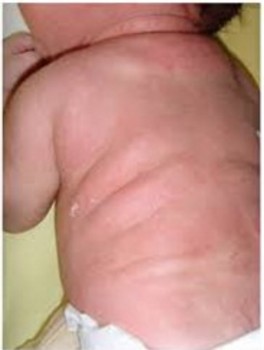
(Subcutaneous Adiponecrosis of the newborn)
Acute hypodermitis developing during the first few days of life. It presents as indurated skin plaques, appearing purple on pale skin or hyperchromic on black skin, and often located on the face, trunk, buttocks and root of the limbs. At histology, under a normal epidermis and derma, there is a lobular panniculitis with areas of eosinophil necrosis of the adipose tissue encompassing optically empty intra-adipocytic radial slits, corresponding to lipid dissolution and crystallization.
At-risk situations include fetal macrosomia (diabetic mother), perinatal asphyxia (circular cord), severe hypothermia (it follows 1 % of therapeutic hypothermia) and tissue trauma occurring during instrumental manoeuvers or during neonatal resuscitation. The evolution is generally benign, but severe hypercalcemia can be associated , le&ading sometimes to life-threatening complications: dehydration, high blood pressure, rhythm disorders (short, then long QT, BAV, enlargement of the QRS), convulsions, nephrocalcinosis, polyuria This hypercalcemia can persist for several weeks after the disappearance of these subcutaneous lesions.
Treatment of hypercalcemia:
- promote calciuresis: hyperhydration with a glucose-enriched saline solution, furosemide 1 mg/kg/8h
- calcitonin 4-8 U/kg/12h
- pamidronate 0.25-0.5 mg/kg
Anesthetic implications:
check the blood electrolytes; in case of polyuria, check the urinary electrolytes to adapt hydroelectrolytic intakes. In case of hypercalcemia: increase IV intakes (1.5 x basal needs) and avoid IV solutions containing calcium.
References :
- Stefanko NS, Drolet BA.
Subcutaneous fat necrosis of the newborn and associated hypercalcemiaĀ: a systematic review of the literature.
Pediatr Dermatol 2019Ā; 36Ā:24-30.
- Matar MM.
A case report and anesthetic considerations in subcutaneous fat necrosis of the newborn.
A&A Practice 2021Ā; 15Ā: e01422
Updated: March 2021